Home / Healthcare Professional / Wilson’s Disease

Diseases

Click here to view more Diseases

Wilson’s Disease

Wilson's disease (WD), also known as hepatolenticular degeneration, is an autosomal recessive disorder characterized by impaired hepatic copper transport and subsequent copper accumulation in various tissues, particularly the liver, and brain.

 
“Several studies indicate that India has a higher prevalence of WD compared to Western countries.”
 

The exact prevalence of Wilson's disease in India remains uncertain due to underdiagnosis and a lack of population-based studies. However, several studies indicate that India has a higher prevalence of WD compared to Western countries. The high frequency of consanguineous marriages and the increased occurrence of WD in certain communities contribute to the disease burden in India. A study conducted by Shalimar et al. in 2019 reported the prevalence of Wilson's disease to be approximately 7.4 per million population in northern India, which is higher than previously estimated. Another study by Sood et al. in 2020 observed a higher prevalence of Wilson's disease (16.6 per million) in the northern Indian state of Punjab. These findings highlight the need for further research and comprehensive nationwide studies to ascertain the true prevalence of Wilson's disease across different regions in India.

Wilson's disease is characterized by impaired copper transport and metabolism in the body. the complex biochemical pathways and pathophysiology of Wilson's disease are illustrated below :

  • Copper from the diet is absorbed in the intestines.
  • ATP7A, a copper transporter protein, facilitates the transport of copper into the bloodstream.
  • Copper is carried in the bloodstream to the liver, where it is taken up by hepatocytes.
  • Inside the hepatocytes, ATP7B, another copper transporter protein, plays a crucial role in transporting copper from the cytoplasm into the Golgi apparatus.
  • In the Golgi apparatus, copper is incorporated into ceruloplasmin, a copper-binding protein.
  • Ceruloplasmin, with copper bound to it, is released into the bloodstream.
  • Copper is then distributed to various tissues and organs where it is required for normal cellular functions.
Pathophysiology

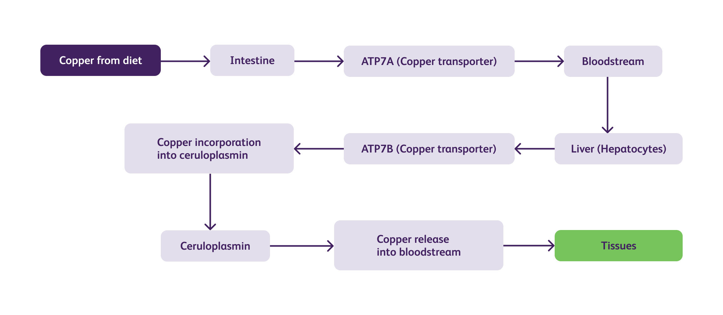
In Wilson's disease, mutations in the ATP7B gene impair the function of the ATPase copper-transporting beta (ATP7B) protein, leading to defective copper transport. As a result, copper accumulates within the liver and is not adequately incorporated into ceruloplasmin, resulting in low levels of circulating ceruloplasmin. The excess copper released into the bloodstream is then deposited in various tissues, including the liver, brain, kidney, and cornea, causing tissue damage and dysfunction.

    The clinical manifestations of Wilson's disease are highly variable and can involve multiple systems.

  • Hepatitis: Most patients present with hepatic symptoms such as hepatomegaly, jaundice, and abnormal liver function tests.
  • Cirrhosis: Prolonged copper accumulation can lead to progressive liver damage, resulting in cirrhosis.
  • Acute Liver Failure: In severe cases, acute liver failure can occur, necessitating urgent medical intervention.

  • Movement Disorders: Tremors, dystonia (abnormal muscle contractions), and dysarthria (difficulty in articulating words).
  • Psychiatric Symptoms: Behavioural changes, personality disorders, and depression.
  • Neurological Decline: Cognitive impairment, difficulty with coordination, and seizures.
  • Parkinsonism: Parkinson's disease-like symptoms such as bradykinesia (slowness of movement) and rigidity.
  • Kayser-Fleischer rings: a characteristic brownish ring around the cornea due to copper deposition.

  • Renal Involvement: Copper build-up in the kidneys can cause renal tubular dysfunction.
  • Haematological Abnormalities: Wilson's Disease may present with low platelet count and anaemia.
  • Osteoporosis: Copper imbalances can affect bone metabolism, leading to osteoporosis.
 

Management

The management of Wilson's disease involves two primary goals: reducing copper accumulation and preventing its toxic effects.


Dietary Modifications

  • Low Copper Diet: Restricting dietary intake of copper-rich foods such as liver, shellfish, mushrooms, and chocolate.
  • Zinc Supplementation: Oral zinc salts inhibit copper absorption in the intestines.
  • Copper-Chelating Medications:
  • D-Penicillamine:

    Enhances urinary copper excretion by forming a complex with copper.

  • Trientine:

    Similar to D-penicillamine, it enhances urinary copper excretion but with fewer side effects than D-penicillamine and is advised in neuro-Wilson’s disease.
    (For more information click here)

  • Liver Transplantation:

    Considered in cases of severe liver damage or failure that do not respond to medical treatment.


  • Aspect D-penicillamine Trientine Tetrahydrochloride
    Dose Initial: 250 mg/day Initial: 750-1,500 mg/day in divided doses
    Gradually increased to 1-1.5 g/day Gradually increased to 750-2,250 mg/day (divided into 2-3 doses) depends on patient's response
    Contraindications Hypersensitivity to penicillamine Hypersensitivity to trientine
    Pregnancy and breastfeeding Pregnancy and breastfeeding
    Neurological Deterioration Has been reported, and treatment should be promptly stopped if any signs of worsening neurological symptoms occur Not reported
    Adverse Effects

    - Rash, fever, proteinuria

    - Hematological abnormalities

    - Nephrotoxicity

    - Autoimmune disorders

    - Neurological symptoms

    - Gastrointestinal disturbances

    - Neurological symptoms (rare)

    - Hematological abnormalities

    - Nephrotoxicity

    - Autoimmune disorders

    - Neurological symptoms

    - Dermatological reactions

    - Myasthenia gravis (rare)

    Contraindicated drugs/Drug-Drug Interactions

    Gold compounds

    Antimalarials

    Cytotoxic drugs

    Aluminum-containing drugs

    Iron salts

    Magnesium salts

    Zinc salts

    Calcium salts

    Nonsteroidal anti-inflammatory drugs (NSAIDs)

    Tetracyclines

    Captopril

    Nitrofurantoin

    Methyldopa

    Anticoagulants

    Digitalis glycosides

    Meclofenamate

    Sulfinpyrazone

    Oral penicillins

    Oral cephalosporins

    Aluminum-containing drugs

    Iron salts

    Magnesium salts

    *Zinc salts

    Calcium salts

    Nonsteroidal anti-inflammatory drugs (NSAIDs)

    Captopril

    Nitrofurantoin

    Methyldopa

    Anticoagulants

    Digitalis glycosides

    Meclofenamate

    Sulfinpyrazone

    penicillins

    Oral cephalosporins

    *Zinc supplements: Zinc supplements may decrease the absorption of Trientine. These medications should be administered at least two hours apart. (Reference: BNF, Trientine Tetrahydrochloride monograph).


    Dietary Modifications

    Low Copper Diet: Restricting dietary intake of copper-rich foods such as liver, shellfish, mushrooms, and chocolate.


New Innovations

Several innovative approaches have emerged for the management of Wilson's disease. Novel copper chelators, such as tetrathiomolybdate, have shown promising results in reducing copper overload. Gene therapy and RNA interference techniques hold potential for targeted gene correction and silencing, respectively. Additionally, advancements in imaging techniques, such as molecular imaging, may aid in early diagnosis and monitoring of treatment response.


Challenges in Management and Diagnosis

Wilson's disease typically manifests between the ages of 5 and 35, with most cases presenting in adolescence or early adulthood. In India, the age of onset appears to be slightly earlier compared to Western populations, with a peak incidence in the second decade of life. This younger age of presentation poses unique challenges in terms of diagnosis and management, as symptoms can be easily mistaken for other common conditions in this age group. Despite advancements, challenges persist in managing and diagnosing Wilson's disease in India. Limited awareness among healthcare professionals and the general population often leads to delayed or missed diagnoses. Inadequate access to specialized diagnostic facilities and genetic testing further hinders accurate identification of the disease. Long-term treatment adherence and monitoring pose additional challenges, necessitating comprehensive multidisciplinary care.


“In India, the age of onset of Wilson’s disease appears to be slightly earlier compared to Western populations, with a peak incidence in the second decade of life.”

Future Directions

Future research in Wilson's disease should focus on improving diagnostic algorithms, particularly in resource-limited settings. Efforts to raise awareness among healthcare professionals and the public are crucial for early detection and intervention. Collaboration between clinicians, researchers, and policy-makers is necessary to establish national registries, facilitate genetic testing, and develop tailored management guidelines. Continued exploration of targeted therapies and gene-based interventions holds promise for personalized treatment approaches.

References

  • Dhawan A, Hathi P. Wilson disease in children: current status and future directions. World J Hepatol. 2015;7(3):582-591.
  • Sheth KA, Sachdeva S, Shah ND, Sheth FJ. Wilson's disease: neurological perspectives. Neurol India. 2018;66(6):1716-1722.
  • Aggarwal A, Bhatt M. Wilson disease: challenges and solutions. Ann Indian Acad Neurol. 2019;22(2):170-177.
  • Walshe JM. Wilson's disease: new insights into pathogenesis, diagnosis, and treatment. Can J Gastroenterol. 2008;22(10):825-830.
  • Weiss KH, Stremmel W. Evolution in understanding the pathophysiological basis of Wilson disease and its treatment. Handb Clin Neurol. 2017;142:33-48.
  • Shalimar, Kumar M, Lal R, et al. Prevalence of Wilson's disease in first-degree relatives of index patients in North India: a descriptive study. J Clin Exp Hepatol. 2019;9(4):460-466.
  • Sood R, Prasad R, Bhardwaj P, et al. High prevalence of Wilson's disease in Punjab, north-west India: need for nationwide screening. Lancet Gastroenterol Hepatol. 2020;5(7):582-590.
  • Gupta, A., & Chattopadhyay, I. (2014). Recent advances on the roles of molecular chaperones in protein-misfolding diseases. Diseases, 2(4), 356-381.
  • Gitlin, J. D. (2003). Wilson disease. Gastroenterology, 125(6), 1868-1877.
  • Roberts EA, Schilsky ML. Diagnosis and treatment of Wilson disease: an update. Hepatology. 2008;47(6):2089-2111.
  • European Association for the Study of the Liver. EASL clinical practice guidelines: Wilson’s disease. J Hepatol. 2012;56(3):671-685.
  • Ferenci P, et al. Wilson's disease: diagnostic tests, treatment options, and management strategies. Hepatology. 2017;67(5):2082-2099.
  • Bandmann O, Weiss KH, Kaler SG. Wilson's disease and other neurological copper disorders. Lancet Neurol. 2015;14(1):103-113.
  • Członkowska A, Litwin T, Dusek P, Ferenci P, Lutsenko S, Medici V. Wilson disease. Nat Rev Dis Primers. 2018;4(1):21. doi:10.1038/s41572-018-0021-8
  • Ala A, Walker AP, Ashkan K, et al. Wilson's disease. Lancet. 2007;369(9559):397-408. doi:10.1016/S0140-6736(07)60196-2
  • Brewer GJ. Wilson disease: update. Curr Opin Gastroenterol. 2013;29(3):245-255. doi:10.1097/MOG.0b013e32835f5610
  • Aggarwal A, Aggarwal N, Nagral A, Jankharia G, Bhatt M. Wilson disease: clinical spectrum and molecular genetic analysis of ATP7B in 103 Indian patients. J Gastroenterol Hepatol. 2006;21(2):384-389.
  • Prashanth LK, Taly AB, Sinha S, et al. Wilson's disease: diagnostic errors and clinical implications. J Neurol Neurosurg Psychiatry. 2004;75(6):907-909.
  • Merle U, Schaefer M, Ferenci P, et al. Clinical presentation, diagnosis, and long-term outcome of Wilson's disease: a cohort study. Gut. 2007;56(1):115-120.
  • Sankhyan N, Mehta S, Sankhyan A, Singhi P. Wilson disease in children and adolescents: 99mTc-DMPS scintigraphy for presymptomatic diagnosis. Pediatr Neurol. 2013;49(5):312-317.
  • Saroli PC, Schilsky ML. Clinical practice guidelines in Wilson disease. Ann Transl Med. 2019;7(2):S65.
  • Socha P, Janczyk W, Dhawan A, et al. Wilson’s disease in children: a position paper by the Hepatology Committee of the European Society for Paediatric Gastroenterology, Hepatology and Nutrition. J Pediatr Gastroenterol Nutr. 2018;66(2):334-344.
  • Nagral A, Sarma MS, Matthai J, et al. Wilson’s disease: clinical practice guidelines of the Indian National Association for Study of the Liver, the Indian Society of Pediatric Gastroenterology, Hepatology and Nutrition, and the Movement Disorders Society of India. J Clin Exp Hepatol. 2019;9(1):74-98.
  • Ferenci P. Diagnosis of Wilson disease. Handb Clin Neurol. 2017;142:171-180.
  • D-penicillamine:British National Formulary (BNF) - Penicillamine. Accessed on May 25, 2023. Available at: https://bnf.nice.org.uk/drug/penicillamine.html.
  • Lexicomp Online. Penicillamine. Accessed on May 25, 2023.
  • Trientine Tetrahydrochloride: British National Formulary (BNF) - Trientine Hydrochloride. Accessed on May 25, 2023. Available at: https://bnf.nice.org.uk/drug/trientine-hydrochloride.html
  • Lexicomp Online. Trientine. Accessed on May 25, 2023.